Moles: When They're Harmless and When To Get One Checked 

What are moles and why do we get them?

A mole, medically known as a nevus, is a small growth on the skin that develops when pigment-producing cells called melanocytes grow in a cluster instead of spreading evenly. Most common moles are round, flat or slightly raised, brown or black spots less than 5mm wide, heavily influenced by genetics, sun exposure, and hormonal changes.

The science behind moles

A common mole is a small, typically brown or black growth on the skin. These spots form when melanocytes clump together. Most common moles have a smooth surface and are less than 5mm wide. Genetics play a large role in how many moles you have. Sun exposure during childhood and hormonal changes, such as those during puberty or pregnancy, can also cause them to develop or darken.

New moles in adulthood

It is completely normal for new moles to appear until you are around 40 years old. Existing moles can also change slightly over time, perhaps getting a little darker or lighter as your hormone levels shift. However, any rapidly changing, unusually shaped, or very dark new mole that appears in adulthood warrants a medical assessment. Most stay harmless throughout life, but changes in symmetry or colour are reasons to speak to a doctor, as advised by the Whittington Health NHS Trust.
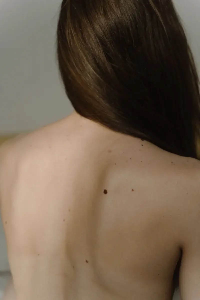
moles-on-back-skin-check

Common types of moles

Common types of moles include benign acquired moles, atypical moles,  and congenital moles, which all share predictable characteristics setting them apart from skin cancer. 

Common acquired moles

A standard, benign nevus is usually small, flat or slightly raised, and uniform in colour. They tend to have a symmetrical shape with smooth, clearly defined borders. The colour is even throughout, usually a single shade of brown, black, or tan.

Atypical moles

Atypical moles are benign growths that can look remarkably similar to melanoma. They are often larger than common moles, have irregular borders, and may feature a mix of colours like pink, brown, and tan. While they are harmless themselves, having a high number of atypical moles increases your risk of developing melanoma. The UKHSA advises that people with many atypical moles should seek medical advice about any suspicious changes to help manage this risk.

Congenital moles

Congenital moles are present from birth. They can range from very small to very large spots covering a significant area of skin. Most are harmless, but very large congenital moles carry a slightly higher risk of developing into skin cancer later in life and should be monitored.

Other common spots

Not every brown mark on your skin is a mole. Freckles are small, flat spots that often fade in the winter. Sun spots, also known as lentigines, are larger flat brown marks that appear on sun-exposed areas as we age. Seborrhoeic keratoses are harmless, wart-like growths that look like they have been stuck onto the skin.

Feature 

Common Mole 

Freckle 

Sun Spot 

Seborrhoeic Keratosis 

Onset 
Childhood to age 40 

Early childhood 

Adulthood 

Over 40 

Size 
Usually under 6mm 

Very small (1 to 2mm) 

Can be over 6mm 

Varies, can be large 

Feel

Flat or slightly raised

Completely flat 

Flat

Raised, crusty, rough 

Colour

Even brown, black, tan 

Light brown 

Light to dark brown 

Brown, black, or pale 

Cancer risk

Low, but requires monitoring 

None

None

None

How to check your moles

To check your moles, perform a full-body skin self-examination in a well-lit room using the ABCDE method to catch any suspicious changes early.

The self-exam process

The best way to check your skin is in a well-lit room after a bath or shower. Use a full-length mirror for your body and a small hand mirror to check hard-to-see areas like your back. Look closely at all areas of your skin. Do not forget to check your scalp, the spaces between your fingers and toes, the palms of your hands, the soles of your feet, and under your nails. It is helpful to take photographs of any moles you want to track, placing a ruler next to the spot to record its exact size. The Leeds Teaching Hospitals NHS Trust recommends checking your skin monthly to become familiar with your moles.

The ABCDE method

The ABCDE checklist is a universal tool used by doctors to distinguish a normal mole from a potential melanoma. The nidirect guidance states that any mole showing these signs should be checked by a professional.
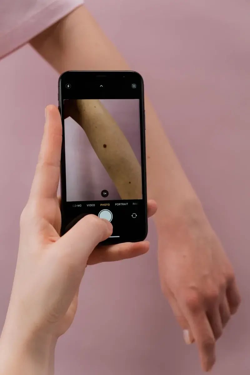
photographing-mole-on-arm-phone

Sign

Meaning

What to look for

A

Asymmetry 

The edges are irregular, ragged, notched, or blurred. 

B

Border 

The colour is uneven, with different shades of brown, black, pink, red, white, or blue. 

C 

Colour 

The colour is uneven, with different shades of brown, black, pink, red, white, or blue. 

D

Diameter 

The spot is larger than 6mm across, roughly the size of a pencil eraser 

E

Evolution 

The mole is changing in size, shape, colour, or height over time. 

The ugly duckling sign

Another very useful concept is the ugly duckling sign. Most of your moles will look relatively similar to each other. If one mole stands out and looks completely different from the rest on your body, it requires medical attention. According to the NHS, you should see a GP urgently if a mole becomes itchy, painful, crusty, or bleeds.

Seeing a doctor about a mole

The clinical process for evaluating a mole involves a careful physical examination followed by a specialist review if there are any signs of concern.

Your GP appointment

When you speak to a GP, they will first take a medical history. They will ask when you first noticed the mole, if it has changed, and whether you have a family history of skin cancer. They will then examine the spot closely. In some cases, a GP may use a dermatoscope, which is a special magnifying lens with a bright light, to look at the pigment patterns beneath the skin surface.

Our online GPs can assess your symptoms in a video appointment and advise you on the right next steps, including whether you need to be seen in person for a physical examination. If a GP suspects a serious issue, they will refer you to a specialist. Under NICE guidance, patients with suspected melanoma are referred urgently to be seen within two weeks.

Visiting a dermatologist

A dermatologist will examine your skin using a high-powered dermatoscope. If they feel the mole needs further investigation, they will perform a biopsy. During a biopsy, the doctor uses a local anaesthetic to numb the area. They then remove a small sample of the mole, or the entire lesion, and send it to a laboratory for testing. This is a quick, minor procedure that causes very little discomfort.

Understanding your results

Your biopsy results will confirm if the mole is benign (harmless), atypical (requiring monitoring or removal), or malignant (cancerous). It is completely normal to feel anxious while waiting for results. If the mole is diagnosed as a melanoma, catching it early leads to a very high cure rate. Most early-stage skin cancers are treated successfully with minor surgery. If you need further support, you can read more about skin cancer treatment.

Mole removal options

The method used to remove a mole depends entirely on whether it poses a health risk or if it is simply causing cosmetic concern or physical irritation.

Medical mole removal

If a mole is diagnosed as skin cancer, it must be removed. The standard procedure is a wide local excision. A surgeon will remove the cancerous tissue along with a margin of healthy skin around it to ensure no abnormal cells are left behind. This is a necessary medical treatment covered by the NHS.

Cosmetic mole removal

Many people choose to remove benign moles because they find them bothersome. For example, a raised mole might catch on clothing or get nicked while shaving. Because this is not medically necessary, cosmetic removal is normally done privately and incurs a cost.

Common methods include a shave excision (slicing the mole off flush with the skin), a punch excision (using a circular tool to remove deeper moles), or laser removal. Every form of mole removal carries a risk of scarring, so it is important to discuss the likely outcome with your clinician.

Avoiding at-home removal

You must never attempt to remove a mole yourself. Do-it-yourself methods like freezing kits, removal pens, or burning creams are highly dangerous and ineffective. At-home removal can cause severe scarring and serious infections. Most critically, these methods carry a terrible risk of leaving cancerous cells behind or destroying a melanoma without it ever being properly diagnosed by a laboratory.

Frequently asked questions

Here are direct answers to the most common questions about how moles look, feel, and change over time.

A hairy mole is not less likely to be cancerous. There is a common myth that hairy moles are always safe, but the presence of hair does not indicate whether a mole is benign or malignant. Skin cancer can develop in any mole, whether it has hair growing from it or not, so you should monitor them all equally.

Having moles on the soles of your feet or palms means you need to monitor them carefully for changes. Acral lentiginous melanoma is a rare type of skin cancer that develops on the palms, soles, or under the nails. The NHS notes that this specific type of melanoma is more common in people with darker skin, making regular checks essential.

Mole mapping is a clinical surveillance programme that uses high-resolution photography to track skin changes, but most people do not need it. According to NICE guidance, such intensive monitoring is recommended primarily for high-risk individuals. This includes those with a strong family history of melanoma or a very large number of atypical moles.

Moles can get itchy or bleed sometimes because they are irritated by dry skin or tight clothing. However, persistent itching or bleeding is a well-known red flag for skin cancer. The Leeds Teaching Hospitals NHS Trust advises that any mole showing unexplained changes like bleeding or crusting must be reviewed by a doctor.

A benign mole can indeed turn into a cancerous one over time. While many melanomas appear as entirely new spots on the skin, a pre-existing, harmless mole can change and become cancerous. The UKHSA warns that changing moles are a key risk factor, which is why the 'Evolution' step in the ABCDE checklist is so crucial.

See a UK GP today

Skip the wait — book an online appointment and get expert advice from a UK-registered clinician in as little as 2 hours. 

Related Topics